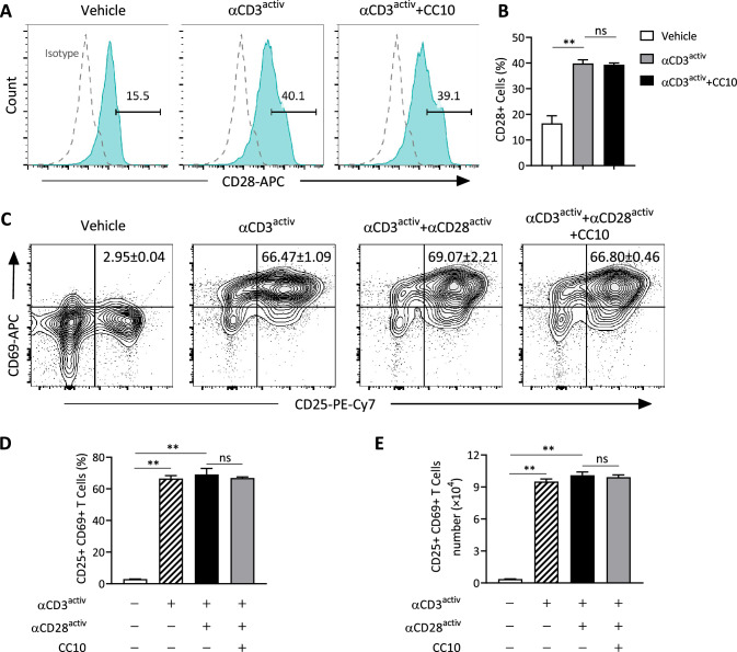
Fig. 4.
CC10 did not directly affect T cell activity. A, B Naïve mouse CD3+ T cells, activated with anti-CD3 mAb, were treated with CC10 (1 μg/mL) for 48 h. Flow cytometry was used to assess CD28 expression (A) and quantify CD28+ cells (B). C T cells, stimulated with immobilized anti-CD3 mAb alone or in combination with soluble anti-CD28 mAb, and CC10 (1 μg/mL) for 48 h, were analyzed for CD25 and CD69 levels via flow cytometry. D, E Quantitative analysis of CD25+CD69+ T cells was conducted. Representing three experiments, data are mean ± SEM. P values derived from one-way ANOVA with Tukey–Kramer test. *P < 0.05, **P < 0.01, ns not significant

