Fig. 3.
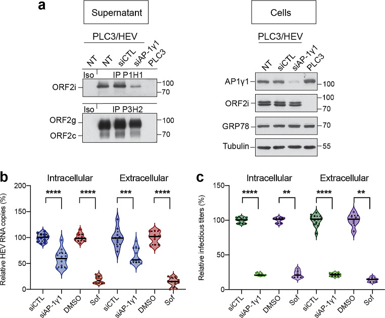
AP-1γ1 silencing affects viral RNA secretion and particle production. (a) Supernatants and cell lysates of non-transfected PLC3/HEV (PLC3/HEV/NT), PLC3/HEV/siCTL, PLC3/HEV/siAP-1γ1 or PLC3 cells were generated 3 days after siRNA transfection. In supernatants, ORF2i and ORF2g/c proteins were immunoprecipitated using anti-ORF2i P1H1 or anti-ORF2i/g/c P3H2 antibodies, respectively. An irrelevant mouse IgG antibody was used as an isotype control (Iso). ORF2 proteins were detected by WB using the 1E6 antibody. In cell lysates, silencing of AP-1γ1 was controlled by WB using a rabbit anti-AP-1γ1 antibody. ORF2i protein was detected using the 1E6 antibody. GRP78 and Tubulin proteins were detected using a rat anti-GRP78 antibody and a mouse anti-β-Tubulin antibody, respectively. (b) HEV RNA quantification in PLC3/HEV/siCTL, PLC3/HEV/siAP-1γ1, PLC3/HEV/DMSO, PLC3/HEV/Sofosbuvir-20µM or PLC3 cells after 3 days of transfection/treatment. Extracellular and intracellular viral RNAs were quantified by RT-qPCR. Titers were adjusted to 100% for siCTL/DMSO-treated cells. PLC3/HEV/Sofosbuvir-20µM cells were used as a positive control for replication inhibition. Values are from four independent experiments. Mann-Whitney test, ***p < 0.001, ****p < 0.0001. (c) Infectious titer determination in PLC3/HEV/siCTL, PLC3/HEV/siAP-1γ1, PLC3/HEV/DMSO, PLC3/HEV/Sofosbuvir-20µM or PLC3 cells after 3 days of transfection/treatment. Extracellular and intracellular viral particles were used to infect naïve Huh-7.5 cells for 3 days. Cells were next processed for indirect immunofluorescence. ORF2-positive cells were counted and each positive cell focus was considered as one FFU. Titers were adjusted to 100% for siCTL/DMSO-treated cells. PLC3/HEV/Sofosbuvir-20µM cells were used as a positive control for infectious titers inhibition. Values are from four independent experiments. Mann-Whitney test, **p < 0.01, ****p < 0.0001

