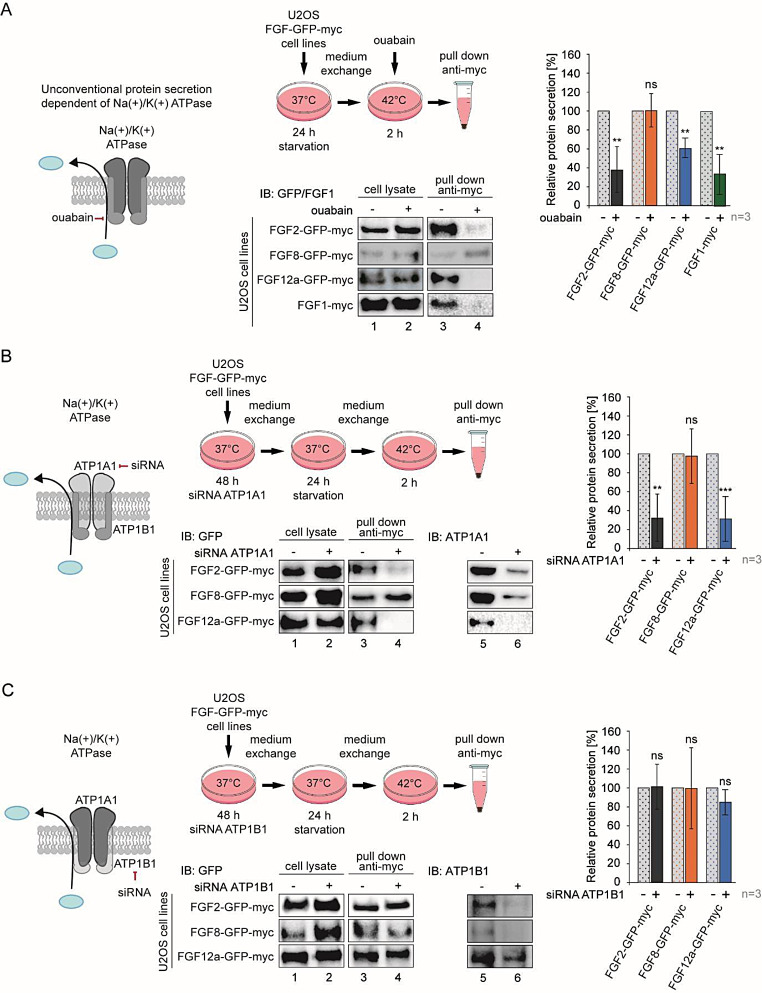
Fig. 2.
The interaction between FGF12a and ATP1A1 is crucial for FGF12a secretion. a U2OS-FGF2-GFP-myc, U2OS-FGF8-GFP-myc, U2OS-FGF12a-GFP-myc and U2OS-FGF1-myc cells were serum-starved at 37 °C for 24 h. After the medium exchange, the cells were incubated at 42 °C for 2 h without or with ouabain (10 µM) and the media from above the cells were collected, centrifuged, and incubated with anti-myc tag magnetic beads. Elutions and lysates were analyzed by SDS-PAGE and western blotting using anti-GFP antibody. The graph shows relative protein secretion quantified using densitometric measurements in Fiji software. Student’s t-test was applied for statistical analysis; ns p > 0.05, ** p ≤ 0.01. b and c U2OS-FGF2-GFP-myc, U2OS-FGF8-GFP-myc and U2OS-FGF12a-GFP-myc cells were transfected with siRNA targeting ATP1A1, ATP1B1 or with scramble siRNA (control) and serum-starved at 37 °C for 24 h after 48 h. The media were exchanged and the cells were incubated at 42 °C for 2 h. The media from above the cells were collected, centrifuged and incubated with anti-myc tag magnetic beads. Eluted proteins and lysates were analyzed by SDS-PAGE and western blotting using anti-GFP antibody. The anti-ATP1A1 and anti-ATP1B1 antibodies were used to verify knock-down effectiveness. Graphs show relative protein secretion quantified using densitometric measurements in Fiji software. Student’s t-test was applied for statistical analysis; ns p > 0.05, ** p ≤ 0.01, *** p ≤ 0.001

