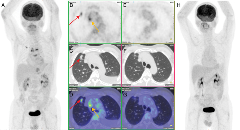
Fig. 2.

Pre- and post-treatment fluorodeoxyglucose positron emission tomography (PET) at first presentation showing complete metabolic response of primary right lung, nodal metastasis and left pleural effusion. ( A ). Pre-therapy maximum intensity projection (MIP) image showing right lung primary tumor and mediastinal nodal metastasis. ( B – D ) Pre-therapy transverse section of PET, computed tomography (CT), and fused images, respectively, showing the lung ( red arrows ) and nodal metastasis ( orange arrows ). ( E – G ) Post-therapy transverse section of PET, CT, and fused images, respectively, showing complete resolution of primary lung and nodal disease. ( H ) Post-therapy MIP image showing complete metabolic response. There was no evidence of FDG-avid local lung carcinoma recurrence or extra peritoneal spread. This led to a change to a 3rd line regimen which consisted of Carboplatin (AUC5), pemetrexed, atezolizumab, bevacizumab. Three months later 18F-FDG PET/CT showed excellent response with complete metabolic remission and no evidence of residual or new FDG-avid malignant or metastatic disease.
