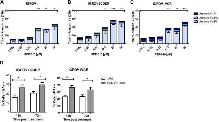
FIGURE 2.
PEP-010 induces apoptosis in OA cells resistant or not to chemotherapies. (A–C) Cells were treated with PEP-010 at the indicated doses for 24 h. Results of treated cells were always compared to those of the untreated control. Analysis of cell death was performed by Annexin V/PI staining and FACS analysis. The results are represented as the total percentage of cells expressing Annexin V. All experiments have been performed in three independent biological replicates. Early apoptosis (Annexin V+/PI-) is represented in light blue; late apoptosis (Annexin V+/PI+) is represented in blue; and necrosis (Annexin V-/PI+) is represented in dark blue. (D) PEP-010 induces DNA damage, as shown by γH2aX expression. Fixed cells were immunostained using an antibody specific for γH2aX as an early marker of DNA damage. The percentage of cells expressing γH2aX was calculated over the total number of cells. Three biological and nine technical replicates and at least 300 cells were analyzed for each biological replicate. Graphs represent the means ± SEM. *, <0.05; **, 0.001 < p < 0.05; and ***, p < 0.001.

