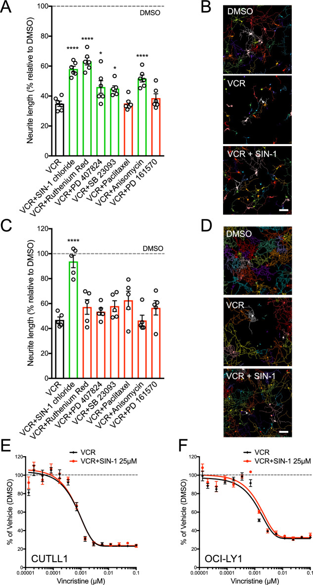
Figure 2.
SIN-1 protects sensory and motor neurons from vincristine-induced neurotoxicity without interfering with vincristine anticancer activity. (A) Effect of 7 lead compounds on pMNs. Neurite length is expressed in percent relative to DMSO (dotted line). Data are presented as Mean ± SEM of 3 independent experiments. One-way ANOVA, repeated measurements with Dunnett’s multiple comparison test (*p ≤ 0.05, ****p ≤ 0.0001 vs. VCR). (B) Representative MetaMorph converted images of GFP positive pMNs in control (0.1% DMSO), 1 nM vincristine (VCR), and 25 µM SIN-1 in the presence of 1 nM VCR (VCR + SIN-1). Scale bar, 100 µM. (C) Effect of 7 lead compounds on DRG neurons. Neurite length is expressed in percent relative to DMSO (dotted line). Data are presented as Mean ± SEM of 3 independent experiments. One-way ANOVA, repeated measurements with Dunnett’s multiple comparison test (*p ≤ 0.05, ****p ≤ 0.0001 vs. VCR). (D) Representative MetaMorph converted images of td-Tomato positive DRG neurons in control (0.1% DMSO), 4 nM vincristine (VCR), and 25 µM SIN-1 in the presence of 4 nM VCR (VCR + SIN-1). Scale bar, 200 µm. (E–F) SIN-1 does not interfere with VCR anticancer activity in cancer lines: CUTTL1 (E) and OCI-LY1 (F). Luminescent cell viability assay based on ATP quantitation in proliferative cells treated with vincristine (VCR, black curves) and vincristine plus SIN-1 (VCR + SIN-1, red curves). VCR doses are expressed in a log scale. Data are presented as Mean ± SEM of 3 independent experiments. See also Figure S2.

