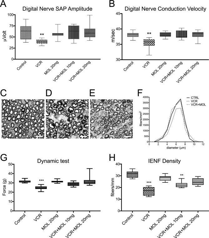
Figure 4.
Molsidomine provides neuroprotection in a rat model of vincristine-induced peripheral neurotoxicity. (A–B) Neurophysiological results obtained at the end of treatment in digital nerves in Sensory Action Potential (SAP) amplitude and sensory and motor conduction velocity. Treatment with 20 mg/kg of MOL restores Sensory Action Potential (SAP) (A) and nerve Conduction Velocity (NCV) (B) of digital nerve altered by VCR to the level of Control. Data are presented as median ± interquartile range (n = 12; One-Way ANOVA with Tukey post-hoc test; **p < 0.01). (C–E) Caudal nerves in control (C), VCR-treated (D), and VCR + MOL-treated (E) rats. Note that VCR alone (D) induces shrinkage of myelinated axons and swelling of extracellular space, and molsidomine cotreatment (E) partially restores the normal ultrastructure of myelinated fibers. Scale bar, 10 µm. (F) Quantification of caudal nerve fiber density in samples from C–E. Molsidomine treatment preserves the nerve density in the caudal nerve. Note that the diameter of axons is preserved in the VCR + MOL group to the level in the Control group, and density is presented as fibers per mm2. Mean ± SEM. One-Way ANOVA on ranks, (G) Treatment with 20 mg/kg of MOL restores a sensitivity threshold to applied mechanical forces decreased by VCR to the level of Control. Data are presented as median ± interquartile range (n = 12; One-Way ANOVA with Tukey post-hoc test; ***p < 0.001). (H) Intraepidermal nerve fiber density (IENF) is preserved in MOL-treated groups compared to VCR. Data are presented as median ± interquartile range (one-way ANOVA and the Tukey–Kramer post-test, n = 3; **p < 0.01, ***p < 0.001). See also Figure S4.

