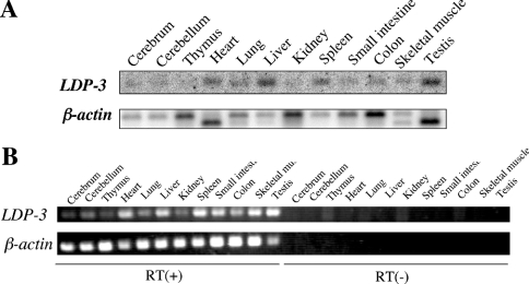
Figure 3. Expression of LDP-3 mRNA in mouse tissues.
(A) Poly(A)+ RNAs (3 μg) obtained from several mouse tissues were separated on a 1% (w/v) agarose gel, transferred to a nitrocellulose membrane, and hybridized with a 32P-labelled mouse LDP-3 cDNA. The membrane was reprobed with 32P-labelled β-actin cDNA. (B) Reverse transcription (RT)–PCR amplification was carried out using specific primer sets for LDP-3 and β-actin.

