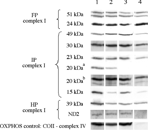
Figure 4. Immunological detection of complex I subunits in mitochondria.
Identical amounts of protein from mitochondrial preparations (8 μg) were loaded. Representative Western-blot signals show the level of each indicated subunit for complexes I and IV in different cell lines. Lane 1, 143B; lane 2, C4T; lane 3, C9T; lane 4, rho°. FP, flavoprotein domain; IP, iron protein domain; HP, hydrophobic part. Assignment of the immunodetected subunits to the three complex I domains was consistent with the results from Hatefi [5]. aDetection with the commercial anti-20 kDa antibody; bDetection with a serum containing antibodies raised against the C-terminal peptide of the 20 kDa subunit. Note that, under our electrophoretic conditions, the 20 and 51 kDa subunits could be detected as doublets by their respective polyclonal antisera. Immunodetection of the same subunits on whole cell samples gave similar results (not shown).

