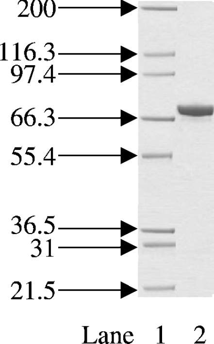
Figure 1. Coomassie-Blue-stained SDS/PAGE gel of the final purified S. aureus DNA ligase.
Protein was expressed and purified as described in the Materials and methods section. The positions and sizes (in kDa) of the of marker proteins are shown on the left. Lane 1, protein size markers; lane 2, 2 μg of total protein. The band at approx. 70 kDa is that of S. aureus DNA ligase.

