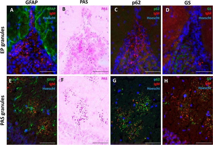
Fig. 2.
Characterization of EP granular structures in mouse brain. When sections were stained with IgM to label the EP granules and with GFAP to stain astrocytes, we observed that EP granules from the fissures showed no relation with astrocytes (A), while the clusters of hippocampal PAS granules were found in areas occupied by astrocytes (E). When staining the sections with PAS, the EP granules were not stained (B), unlike PAS granules, which, as expected, were stained with the PAS stain (F). EP granules were not stained with p62 or GS (C and D, respectively), while PAS granules of the hippocampus were positive to these stainings (G and H, respectively). Sections A, C, D, E, G and H were also stained with Hoechst (cell nuclei, blue). Scale bars: 50 µm

