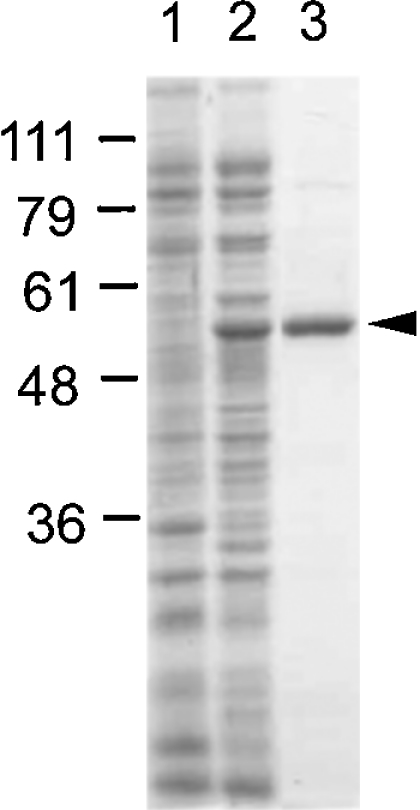
Figure 2. UDP-GlcDH expression and purification.

Bacterial proteins analysed by SDS/PAGE and stained with Coomassie Blue. Standards (in kDa) are shown on the left. Lane 1, 20 μg of soluble protein from E. coli expressing plasmid only; lane 2, 20 μg of soluble protein from E. coli expressing His-tagged UDP-GlcDH; lane 3, 2 μg of UDP-GlcDH purified by cobalt chromatography. A minor contaminant is barely visible at approx. 90 kDa.
