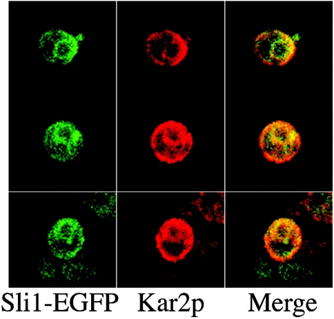
Figure 7. Intracellular localization of Sli1p.
Yeast cells containing YEp351ADH1-Sli1p-EGFP were fixed with formaldehyde, converted into spheroplasts, and permeabilized with 0.1% Triton X-100. They were then stained with anti-Kar2p antibodies. Green indicates the localization of Sli1p–EGFP (left panel) and red indicates the localization of Kar2p (middle panel). In the merged image (right panel), yellow indicates the co-localization of Sli1p–EGFP and Kar2p.

