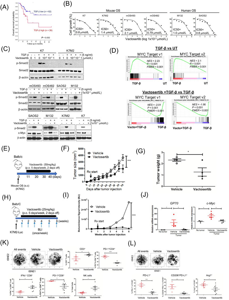
FIGURE 1.

Vactosertib inhibits osteosarcoma cell growth in vitro and in vivo. (A) Kaplan‐Meier overall survival curves of high‐grade osteosarcoma patients and their expression of TGF‐β1 in clinical biopsies. Data were obtained from “R2: Genomics analysis and visualization platform” [http://r2.amc.nl]. Datasets provided by Kuijer (n = 88). The red line indicates high expression of TGF‐β1 (n = 26), while the blue line indicates low expression of TGF‐β1 (n = 62). Kaplan‐Meier curves showed worse overall survival rates of osteosarcoma patients with high TGF‐β expression compared to patients with low TGF‐β1 expression (P = 0.032). (B) Effects of vactosertib on osteosarcoma proliferation. Various doses of vactosertib (10×10−3 µmol/L to 10 µmol/L) were incubated with mOS (K7, K7M2, mOS493, and mOS482) or hOS (M132 and SAOS2). Cell growth was quantified over a 4‐day period using the IncuCyte Imaging System. The non‐linear regression (curve fit) equation was calculated using GraphPad prism (n = 5/group). (C) Vactosertib inhibits TGF‐β1 signaling pathway in osteosarcoma cells. Various doses of vactosertib (1×10⁻² µmol/L to 1 µmol/L) were used to treat in K7, K7M2, mOS493, mOS482, SAOS2 and M132 cells 15 minutes before TGF‐β1 (5 ng/ml) treatment. 1 hour after TGF‐β1 treatment, cells were harvested and p‐Smad2, Smad2 and β‐actin expressions were measured by Western blot analysis. TGF‐β1 (5 ng/ml) or TGF‐β1 (5 ng/ml) / vactosertib (Vacto) (1×10⁻¹ µmol/L) were used to treat various osteosarcoma (SAOS2, M132, K7M2, K7) for 24 hours and c‐Myc and β‐actin protein expressions were measured by Western blot analysis. (D) RNA‐sequencing analysis of human osteosarcoma (SAOS2) cells after TGF‐β1 (5 ng/ml), TGF‐β1+ vactosertib (1×10⁻¹ µmol/L) or untreated (UT) for 24 hours. GSEA enrichment plot of c‐Myc target v1 and v2 pathway in TGF‐β1 treatment versus untreated and in TGF‐β1 + Vacto vs TGF‐β1. P values are < 0.001 and FWER is < 0.001 for both analyses. (E‐G) Vactosertib inhibited OS cell growth in vivo. (E) BALB/c mice were inoculated with 1×10⁶ K7M2 (s.c.) on Day 0, and then treated with vehicle (p.o.) or vactosertib (50 mg/kg, p.o. 5 days/week) starting on day 11. (F) tumor sizes were measured by caliper. n = 4, ***P < 0.001 using a two‐way ANOVA between groups followed by post‐hoc Bonferroni's multiple comparison tests. (G) Tumor weight was measured (n = 4). (H‐J) Vactosertib inhibits mouse pOS development in vivo. (H) BALB/c mice were inoculated with 1×10⁶ K7M2‐Luc (i.v.) on day 0, and then treated with vehicle (p.o.) or vactosertib (25 mg/kg p.o. 5 days/week) starting on day 7. (I) BLI was measured once a week. (J) Relative mRNA expression of GP70 and c‐Myc in lung samples of the vehicle or vactosertib‐treated mice on day 42 days after tumor injection compared with that of control lungs of no tumor‐bearing mice. n = 5/group, *P < 0.05 using an unpaired two‐tailed t‐test. (K‐L) BALB/c mice were inoculated with 1×10⁶ K7M2‐Luc (i.v.) on Day 0, and then treated with vehicle (p.o) or vactosertib (50 mg/kg, p.o. 5 days/week) starting on day 28 (4 weeks). Ten weeks after tumor injection, lung samples were collected. (K) FACS was performed and expression of CD3, CD4, CD8, NK (CD49b), PD‐1 and IFNγ was determined by FACS. Unbiased immune cell profiling on 5000 live CD45.2 cells by t‐SNE analysis was performed. tSNE density plots of live cells in vehicle or vactosertib‐treated samples. The frequency of T cell markers by conventional FACS analysis. Vehicle n = 4, vactosertib n = 7 *P < 0.05, ***P < 0.001, using an unpaired two‐tailed t‐test. (L) Examined for expression of F4/80, PD‐L1, CD206 and Arg1 by FACS. tSNE density plots of CD45.2+ cells in vehicle or vactosertib‐treated samples. The frequency of myeloid cell markers by conventional FACS analysis. Vehicle n = 4‐7, vactosertib n = 7‐9, *P < 0.05, **P < 0.01 using an unpaired two‐tailed t‐test. Abbreviations: BLI, bioluminescent imaging; c‐Myc, cellular‐Myelocytomatosis; FACS, fluorescence‐activated cell sorting; hOS: human osteosarcoma; p.o., per os; PD‐1, Programmed death 1; PD‐L1, programmed death‐ligand 1; RNA‐seq, RNA sequence; s.c., subcutaneous; TGF‐β1, Transforming growth factor‐beta 1; tSNE, t‐distributed stochastic neighbor embedding; UT, untreated; Vacto, vactosertib.
