
Figure 4.
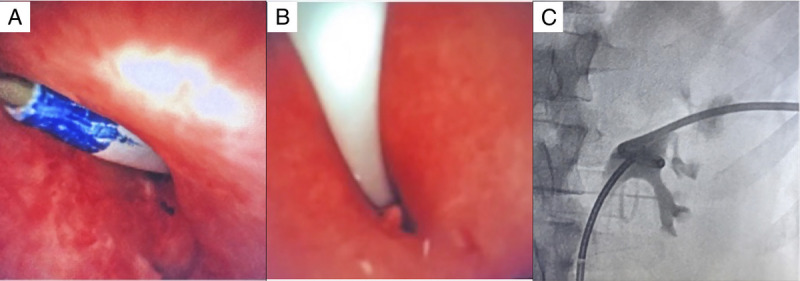
Nephrostomy tube installation. (A) Dilation of the puncture site. (B) Nephrostomy tube installation under flexible endoscope control. (C) Antegrade pyelography after installation of the nephrostomy tube and ureteral stent.
Official websites use .gov
A
.gov website belongs to an official
government organization in the United States.
Secure .gov websites use HTTPS
A lock (
) or https:// means you've safely
connected to the .gov website. Share sensitive
information only on official, secure websites.
Nephrostomy tube installation. (A) Dilation of the puncture site. (B) Nephrostomy tube installation under flexible endoscope control. (C) Antegrade pyelography after installation of the nephrostomy tube and ureteral stent.