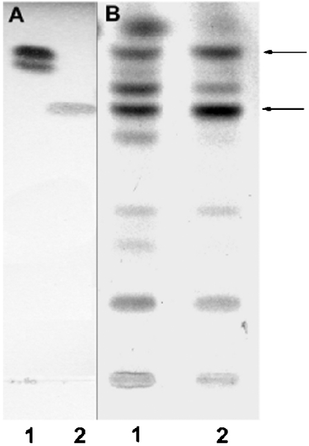
Figure 1. Ceramide biosynthesis by purified mitochondria and MAM in vitro.
Purified mitochondria and MAM were incubated with [3H]sphingosine and unlabelled palmitoyl-CoA under the conditions described in the Experimental section. After purification of the total lipid extract on solid-phase LC-NH2 columns, the fractions corresponding to ceramides (fractions 2 according to [28]) were analysed on silica gel TLC in the solvent system chloroform/methanol (12.5:1, v/v), along with standards. (A) Standards visualized at 150 °C after spraying with copper acetate (3%) in phosphoric acid (8%). Lane 1, ceramides type III; lane 2, phytoceramides. (B) Autoradiogram of fractions 2 eluted from LC-NH2 columns. Lane 1, mitochondria; lane 2, MAM. Arrows indicate the spots that were scraped and re-run in Figure 2.

