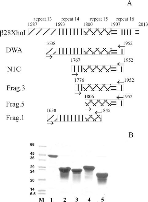
Figure 1. The β-spectrin ankyrin-binding domain and its truncated mutants.
(A) Schematic representation of the ankyrin-binding domain of erythrocyte spectrin and of the location of primers (→) used to amplify appropriate DNA fragments. Clone β 28 Xho was obtained from Dr J.S. Morrow [29]. DWA is the full-length ankyrin-binding domain, and N1C, Frag.1, Frag.3 and Frag.5 denote specific truncated mutants as indicated. (B) Amplified DNA fragments coding for the full-length ankyrin-binding domain of erythrocyte β-spectrin and its truncated mutants obtained by PCR using appropriate primers were cloned into pRSET vector. Recombinant proteins were expressed in a bacterial strain BL-21 expression system using isopropyl β-D-thiogalactoside induction for 3 h at 37 °C, and were purified by immobilized Co2+-affinity chromatography (Clontech). A Coomassie Blue-stained SDS/polyacrylamide gel (10%) electophoretogram of the expressed proteins is shown. Lanes: M, protein markers (molecular masses in kDa are shown to the left); 1, DWA (full-length ankyrin-binding domain); 2, N1C; 3, Frag.3; 4, Frag.1; 5, Frag.5.

