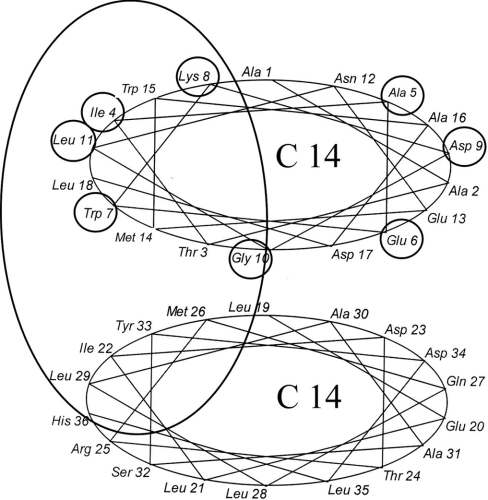
Figure 8. ‘Helical wheel’ representation of the N-terminal part of the erythrocyte β-spectrin ankyrin-binding domain.
The first eight amino acid residues are circled. The vertical ellipse indicates the presumed phospholipid-binding surface. The start of the helical segment is based on data from [40].

