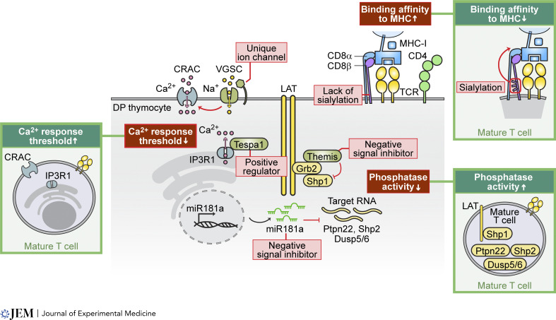
Figure 4.
Regulatory mechanisms for enhanced sensitivity in preselection DP thymocytes. This illustration depicts the three key molecular mechanisms that confer augmented sensitivity in immature DP thymocytes, enabling their activation by weak positively selecting signals that normally cannot activate mature T cells. First, the sialylation pattern on CD8β determines its binding strength to MHC-I molecules, with the lack of sialylation in DP thymocytes increasing their binding affinity. Second, immature DP thymocytes express stage-specific regulatory proteins that lower response thresholds, augmenting calcium influx. For example, the expression of voltage-gated sodium channels (VGSC) promotes sustained calcium responses through calcium release-activated channels (CRAC) in response to weak ligand stimulation. Additionally, the regulatory protein Tespa1 directly interacts with IP3R1 to enhance calcium release from the endoplasmic reticulum. Lastly, elevated expression of negative signal inhibitors, such as Themis and miR-181a, inhibits phosphatase activity, allowing immature DP thymocytes to be activated by weaker TCR stimuli. The illustrations in the green frames highlight the differential mechanisms in mature T cells, emphasizing the unique features of immature DP thymocytes in the middle.

