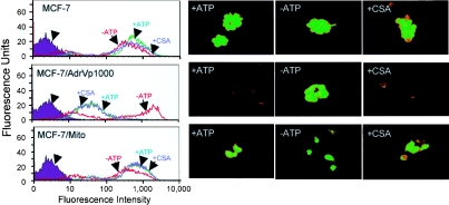
Figure 2. Intracellular uptake of Rh123.
Cells (MCF-7, MCF-7/AdrVp1000 and MCF-7/Mitox) were grown in six-well plates, washed with PBS and incubated in the presence or absence of inhibitors of ATP synthesis or cyclosporine A (CSA), as described in the Materials and methods section. Cells were kept for 30 min at 37 °C before adding Rh123 to each well at a final concentration of 1 μM, with or without 2 μM cyclosporine A (as indicated in the Figure). Left panels: cells were washed and subjected to flow cytometry by FACscan using Rh123 excitation at λ=485 nm. Right panels: cells after incubation were viewed at ×400 magnification using a Nikon TE200 inverted microscope equipped with a fluorescence filter.

