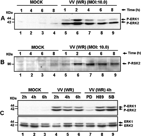
Figure 1. Sustained kinase activation after VV infection.
Cells were serum-starved and then infected with VV at the MOI and times indicated. Western-blot analysis was performed with cell lysates, and 30 μg of protein/lane were fractionated by SDS/PAGE, transferred on to nitrocellulose and then probed with the specific anti-phospho-antibody. Cells were either mock- or VV-infected. (A, B) ERK1/2- and RSK2-activation respectively. (C) Upper panel: PD98059 caused a specific inhibition of ERK1/2 phosphorylation. Cells were either left untreated (lanes 1–6), or preincubated for 30 min with protein kinase inhibitors: 50 μM MEK (PD98059), lane 7; 20 μM PKA (H89), lane 8 or 10 μM p38MAPK (SB203580), lane 9; then mock-infected (lanes 1–3) or VV-infected (WR) at an MOI of 10.0 as indicated. Lower panel: the same blot was re-probed with total ERK as an internal control for protein loading. The position of the phosphoproteins or the molecular-mass standard (kDa) is indicated on the right or on the left respectively. The results were consistently repeated in at least two independent experiments.

