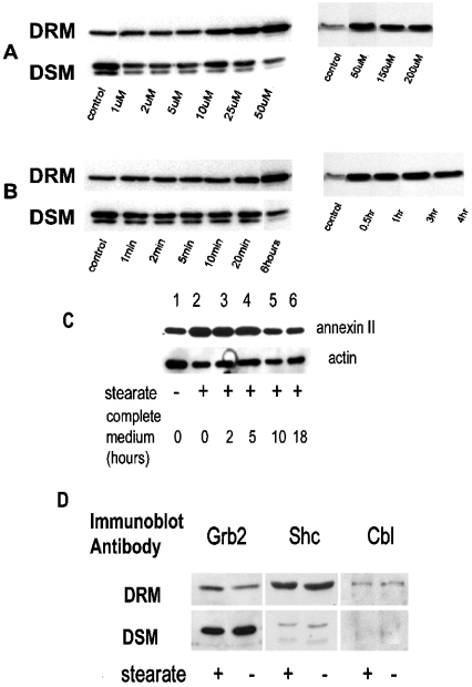
Figure 2. Stearate-induced movement of annexin II to DRM is concentration- and time-dependent, reversible and specific.
Hs578T cells were incubated with stearate at different concentrations as indicated in (A) for 6 h and lysed. DRM and DSM were prepared, resolved by SDS/PAGE and immunoblotted for annexin II as described in the Experimental section. The left and right panels in both (A, B) are two separate experiments. In (B), cells were incubated with 50 μM stearate for the indicated times. In (C), cells were treated with 50 μM stearate for 6 h; then the stearate was removed and cells were incubated with complete media for the times indicated and DRM were prepared as described in the Experimental section. Immunoblot was developed for annexin II and actin as indicated (representative of three separate experiments). Densitometry results of the three experiments demonstrate that the stearate-treated samples at 0 and 2–5 h are increased versus controls (P=0.0217 and P=0.0439 respectively), whereas the 10 h and 18–24 h samples were not different from the controls (P=0.6366 and P=0.8837 respectively). In (D), Hs578T cells were serum-starved 48 h before use and treated with or without 50 μM stearate for 6 h. DSM and DRM were prepared, and proteins were resolved by SDS/PAGE, transferred on to PVDF and immunoblotted as indicated (representative of two separate experiments). Although there appears to be an increase in Grb2 in the DRM in the presence of stearate in comparison with the control, this was not a consistent observation.

