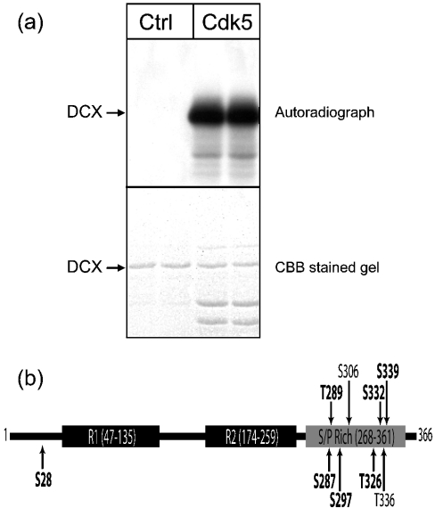
Figure 1. DCX is a substrate for phosphorylation by cdk5 on multiple sites.
(a) In vitro phosphorylation of DCX. GST–DCX was phosphorylated for 10 min at 37 °C in the presence of GST-cdk5/p25. The upper panel shows an autoradiograph, whereas the lower panel shows the Coomassie Brilliant Blue (CBB)-stained gel from the same experiment. DCX was incubated without any protein kinase (lanes 1 and 2) or with cdk5 (lanes 3 and 4). Results are shown in duplicate from one of six experiments. (b) Schematic representation of mouse DCX, indicating the location of R1 and R2 domains and the serine/proline-rich region. Arrows indicate the location of potential cdk5 phosphorylation sites and those identified in the present study are shown in bold text.

