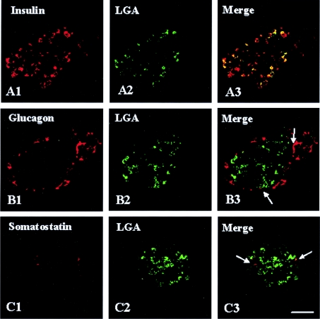
Figure 2. LGA is abundantly expressed in β-cells from rat pancreas.
Double-immunofluorescence labelling with anti-LGA (green) and endocrine cell markers: insulin, glucagon or somatostatin (red). In confocal overlay images, yellow represents co-localization of the two antigens. (A3) LGA and insulin, (B3) LGA and glucagon and (C3) LGA and somatostatin. Note that, whereas most of the insulin-containing cells exhibited a strong LGA immunoreactivity, only a few α- and δ-cells showed a weak, albeit detectable (arrows), immunolabelling. Scale bar, 57 μm.

