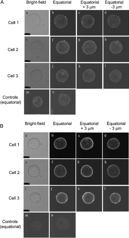
Figure 3. Localization by immunofluorescence of the type I (A) and type II (B) Ins(1,4,5)P3Rs in rat hepatocytes grown for 2 h in primary culture.
The culture of rat hepatocytes, permeabilization and cell fixation, staining with anti-Ins(1,4,5)P3 antibody, and confocal microscopy were performed as described in the Materials and methods section. (A) Cells stained with mouse monoclonal anti-Ins(1,4,5)P3R1 antibody KM1112 and anti-mouse antibody conjugated to Cy3. (B) Cells stained with mouse monoclonal anti-Ins(1,4,5)P3R2 antibody KM1083 and anti-mouse antibody conjugated to Cy3. The results shown are those obtained for one of three experiments employing three separate rat hepatocyte preparations which each gave similar results. The scale bar represents 10 μm. Panels a–d, e–h and i–l each represent a different single cell, showing a bright-field image, an equatorial image, 3 μm above equatorial image (z plane) and 3 μm below equatorial respectively. Panels m and n are controls in which primary antibody has been omitted. All images in (A) and (B) were obtained with the same confocal gain setting.

