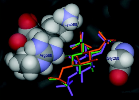
Figure 5. Structure of the Ins(1,4,5)P3R1-binding site based on the X-ray crystal structure of the mouse Ins(1,4,5)P3R1 core binding domain.
The crystallographically observed position of bound Ins(1,4,5)P3 [39] is shown in orange. Molecular docking experiments suggest that Ins(1,4,6)P3 (green) may be a relatively effective mimic of Ins(1,4,5)P3 at the Ins(1,4,5)P3R1-binding site because it can bind in an orientation that allows its phosphate groups to mimic the three phosphate groups of Ins(1,4,5)P3, while its axial 2-hydroxy group is accepted by an open region close to Gly268. However, Ins(1,3,6)P3 (purple) may be prevented from adopting a similar binding mode due to unfavourable steric interactions of its axial 2-hydroxy group with Arg568 and Lys569. Molecular-docking experiments were carried out using the X-ray crystal structure of the Ins(1,4,5)P3-binding core of mouse type 1 InsP3R in complex with Ins(1,4,5)P3 (PDB code 1N4K [39]) according to methods previously described [43]. For clarity, six molecules of water included in the docking experiments, and the hydrogen atoms of Ins(1,4,5)P3, Ins(1,4,6)P3 and Ins(1,3,6)P3 are not shown.

