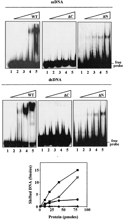
Figure 5. DNA-binding activity of full-sized SsoCdc6-2 and its truncated forms.
The ability of the proteins to bind a 32P-labelled 51-mer synthetic oligonucleotide in ss and ds form was analysed by gel-shift assays, as indicated in the Experimental section. In these experiments, increasing amounts of SsoCdc6-2, ΔC or ΔN were used (10, 20, 40 or 80 pmol). Detection and quantification of the radioactive bands was carried out using a PhosphorImager. A plot of shifted DNA (ss form, open symbols; ds form, closed symbols) against the amount of protein used (full-sized SsoCdc6-2, squares; ΔN, circles; ΔC, triangles) is shown in the bottom panel. Results are mean values of at least three independent experiments.

