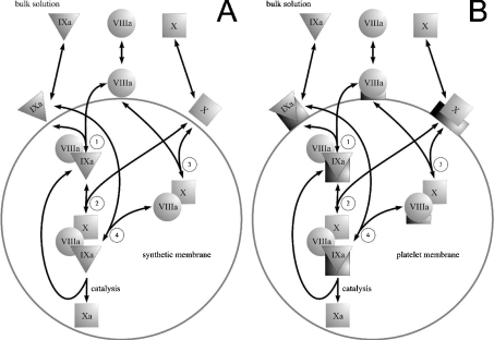
Figure 2. Mechanisms of fX activation by intrinsic tenase used in the model.
(A) Assembly of the intrinsic tenase complex and fX activation on the surface of phospholipid vesicles. fIXa, fVIIIa and fX bind to the membrane from solution. Membrane-bound factors form the fIXa–fVIIIa and fVIIIa–fX complexes (reactions 1 and 3), which subsequently bind fX (reaction 2) and fIXa (reaction 4) respectively to form the final fIXa–fVIIIa–fX complex. (B) Assembly of the intrinsic tenase complex and fX activation on the platelet membrane. fIXa, fVIIIa and fX bind to the membrane from solution; in contrast with (A), each factor binds to its specific site (to illustrate the binding specificity, platelet binding sites for fIXa, fVIIIa and fX are represented as shapes complementary to triangles, circles and squares respectively). fVIIIa and fIXa form intrinsic tenase localized on fIXa receptor, which subsequently binds fX. Platelet-bound fVIIIa can also form a receptor for fX [21], which is followed by the binding of fIXa [22] and the assembly of the final enzyme–cofactor–substrate complex.

