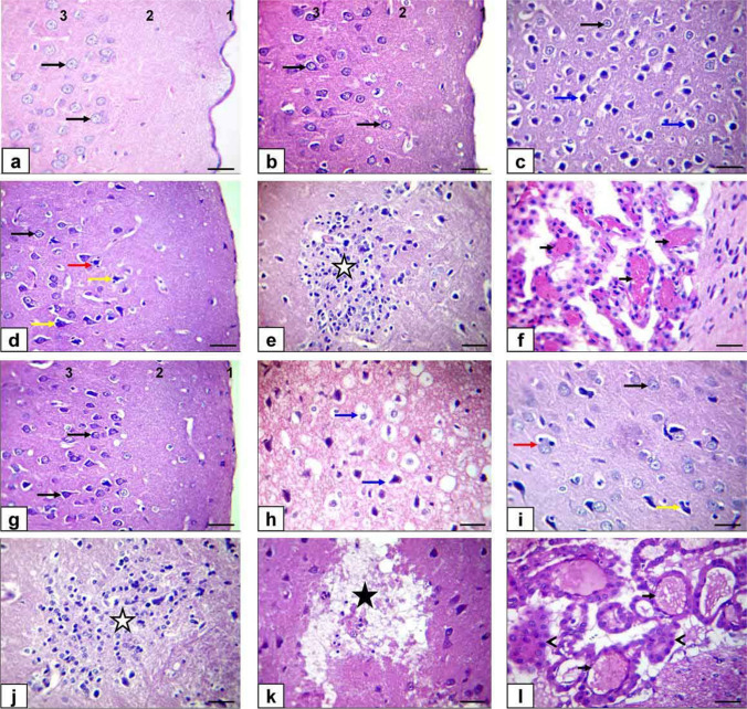
Fig. 6.
Photomicrograph of the cerebrum sections of rats stained by H&E (bar=50μm). a Control rats. b ZnONP-5-treated rats. c, d, e, and (f) ZnONP-10-treated rats. g BZnO-5-treated rats. h, i, j, k, and l BZnO-10-treated rats. Meninges (1), molecular layer (2), external granular layer (3), normal cortical neuron (black arrows), shrunken, pyknotic, and hyper-eosinophilic cytoplasm with widening of neuropil (blue arrows), satellitosis (red arrow) neuronophagia (yellow arrows), gliosis (white star), polioencephalomalacia (black star), and congestion of choroid plexus vasculature (short black arrows) destruction of choroid plexus epithelium (arrowheads)

