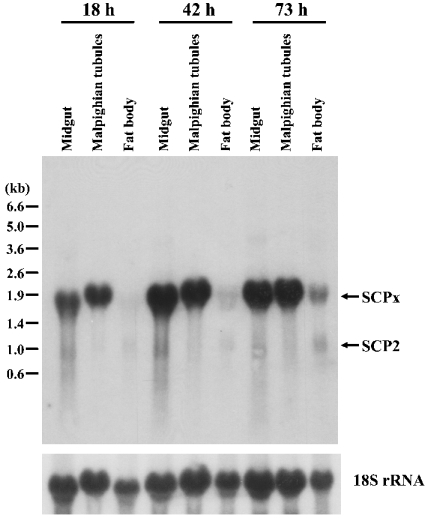
Figure 3. Northern-blot analysis of the tissue distribution and expression of SCPx.
A 10 #x3BC;g aliquot of total RNA from various tissues at different times (top) within the last larval instar was used. The blot was hybridized with a 32P-labelled SCPx cDNA probe corresponding to the whole protein-coding region. The same blot was stripped and reprobed with 18 S rRNA probe to normalize sample loading. The positions of RNA size markers are shown on the left.

