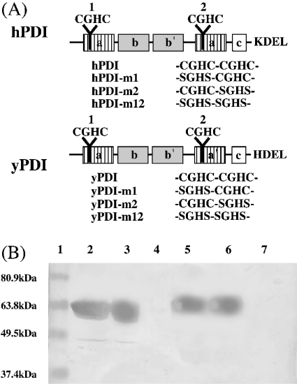
Figure 6. The domain structures of hPDI and yPDI, the sequences of their mutants and recognition of the mutants by the 5E phage antibody by Western blotting.
(A) Domains a and a′ (striped boxes), redox-active Trx domains; domains b and b′ (grey boxes), redox-inactive Trx domains; and domain c (white box), a putative calcium-binding domain. The C-terminal KDEL and HDEL sequences are the possible ER-retention signals. (B) Detection of hPDI and yPDI mutants by the 5E phage antibody by Western blotting. Lane 1, marker proteins; lane 2, hPDI-m1; lane 3, hPDI-m2; lane 4, hPDI-m12; lane 5, yPDI-m1; lane 6, yPDI-m2; lane 7, yPDI-m12. hPDI and yPDI are shown in Figure 2. The 5E phage antibody (1011 pfu/ml) was used with each protein (approx. 5 μg).

