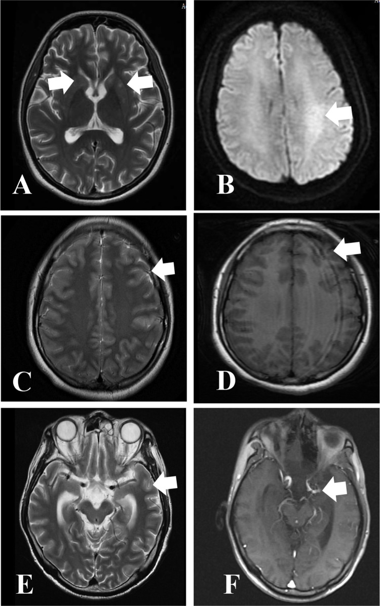
Figure 1.
Magnetic resonance imaging images of selected CNS infections cases. Abnormal signal in bilateral basal ganglia in Case 22 (A) (Arrows: bilateral basal ganglia). Abnormal signals in Left semioval center and parieto-occipital lobe, possible inflammatory lesions in case 15 (B). (Arrows: Left semioval center) Multiple streak-like high signals are seen in the cerebral sulci in case 1 (C), (Arrows: cerebral sulci) Patchy high signal intensity was seen in the left temporal lobe in case 1 (D). (Arrows: left temporal lobe) Linear enhancement on the surface of the brain gyrus in case 11 (E), (Arrows: brain gyrus) thickening and enhancement of the ependyma in the left temporal horn of the ventricle in case 11 (F).(Arrows: Left temporal horn of the ventricle).

