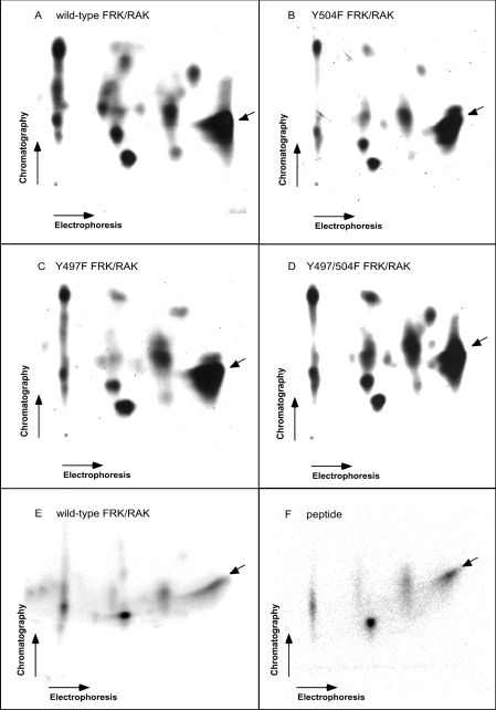
Figure 1. Tryptic peptide fragments of baculovirus-produced FRK/RAK autophosphorylation sites.
Wild-type (A, E), Y504F (B), Y497F (C) and Y497/504F (D) murine FRK/RAK is shown. In (F), the phosphopeptide map of in vitro phosphorylated VDNEDIYESKHEIKKK peptide is shown, of which one position is identical with that of the wild-type FRK/RAK pattern. (E) is identical with (A), except that the experiment was performed at a different time.

