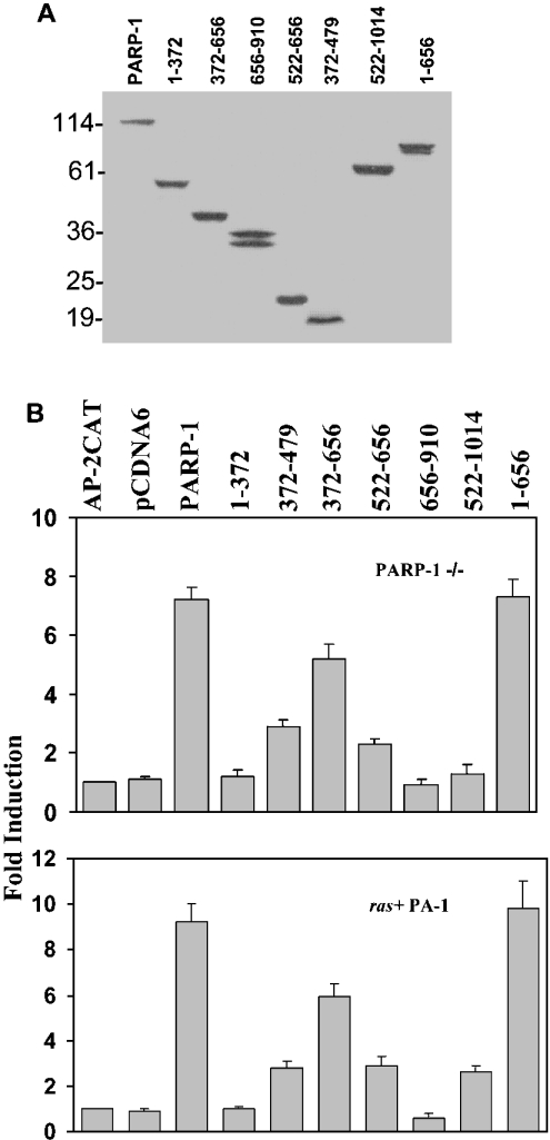
Figure 5. Mapping the regions of PARP-1 that affect AP-2 transcription.
(A) Expression of truncated PARP-1 molecules in transiently transfected PARP-1−/− MEF cells. Expression plasmids (4 μg) of PARP-1 deletion molecules were transfected into these cells. After 48 h, total protein was isolated and 100 μg of protein was resolved on an 18% SDS/polyacrylamide gel, Western-blotted and probed with Myc-Ab. (B) The middle region of PARP-1 enhances AP-2α transcription. Expression plasmids (4 μg) of PARP-1 deletion molecules and 4 μg of 3XAP-2hmtCAT were transiently transfected into PARP-1−/− and ras+ PA-1 cells and the CAT assays were performed. The activity of the reporter plasmid alone is taken as 1 to calculate fold induction. Results are from three independent experiments.

