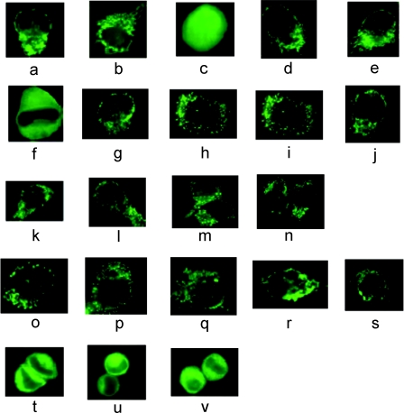
Figure 1. Fluorescence microscopy of HeLa cells transiently expressing EGFP fusion or HA-tagged proteins.
HeLa cells were cultured on coverslips and transfected with 2.5 μg of plasmid DNA. Fluorescence microscopy was used to localize the EGFP constructs. HA-tagged proteins were detected using immunofluorescence. The expressed proteins that lacked a mitochondrial signal were distributed throughout the cell, including the nucleus. The proteins with mitochondrial signals were localized to mitochondria, and are seen as either small dots or long cylinders. The absence of fluorescence in the cytosol and nucleus is indicative of efficient mitochondrial import. The proteins that had only an ER or peroxisomal signal were localized to the ER, appearing as a lacy network of membranes, or the peroxisome, appearing as bright dots. (a) PreOTCsp–EGFP, (b) preAIIsp–EGFP, (c) EGFP, (d) preOTCsp–EGFP-ER, (e) preAIIsp–EGFP-ER, (f) EGFP-ER, (g) preOTCsp–EGFP–SKL, (h) EGFP–SKL, (i) preALDHsp–HA, (j) rhodanese–HA, (k) preOTCsp–HA, (l) ALDH–HA–SKL, (m) OTC–HA–SKL, (n) (Δ2–30)-rhodanese–SKL, (o) preALDHsp–HA–SKL, (p) preALDHsp–HA-ER, (q) preOTCsp–HA-ER, (r) preOTCsp–HA–SKL, (s) rhodanese–HA–SKL, (t) ALDH-ER, (u) OTC-ER, (v) (Δ2–30)-rhodanese-ER.

