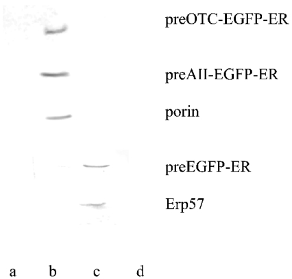
Figure 2. Western blot analysis of preOTCsp–EGFP-ER, preAIIsp–EGFP-ER and EGFP-ER expressed in HeLa cells and after subcellular fractionation.
Subcellular fractionation was described in the Materials and methods section. Different organelles were subjected to Western blot analysis. PreOTCsp–EGFP-ER, preAIIsp–EGFP-ER and EGFP-ER were each transfected into HeLa cells and detected by the anti-EGFP antibody. Human porin was detected using its monoclonal antibody, and served as a mitochondrial marker. Anti-Erp57 antibody was used as an ER-specific marker. Lanes a, b, c and d represent nucleus-, mitochondria-, ER- and cytosol-rich fractions respectively.

