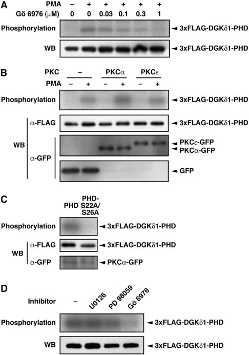
Figure 3. Effects of a cPKC-selective inhibitor, Gö 6976, and PKCα expression on the DGKδ1-PH domain phosphorylation.
(A) COS-7 cells were transfected with a plasmid encoding the 3×FLAG–DGKδ1-PH domain. Cells were starved, 32P-labelled and incubated with Gö 6976 at the indicated concentrations for 30 min before PMA stimulation. Cells were stimulated with 100 nM PMA (+) or 0.1% DMSO (−) for 30 min in the presence of Gö 6976. (B) COS-7 cells were co-transfected with p3×FLAG–CMV–DGKδ1-PH domain and either pEGFP alone, pEGFP–PKCα or pEGFP–PKCε. After starvation and 32P-labelling, cells were incubated for 30 min in the presence of 100 nM PMA (+) or 0.1% DMSO (−). (C) COS-7 cells were co-transfected with pEGFP–PKCα and either p3×FLAG–CMV–DGKδ1-PH domain or -PH domain-S22A/S26A. After starvation and 32P-labelling, cells were incubated for 30 min in the presence of 100 nM PMA. (D) COS-7 cells were transfected with a plasmid encoding the 3×FLAG–DGKδ1-PH domain. Cells were starved, 32P-labelled and incubated with U0126 (50 μM), PD098059 (100 μM) or Gö 6976 (1 μM) for 30 min before PMA stimulation. Cells were treated with 100 nM PMA for 30 min in the presence of the inhibitors. FLAG-tagged proteins were immunoprecipitated with anti-FLAG antibody and analysed by SDS/PAGE. The radioactive signal was visualized by a BAS1800 Bio-Image Analyzer (A–D, top panels), and the 3×FLAG-tagged proteins immunoprecipitated (A–D) and the GFP-fusion proteins in cell lysates (B and C) were analysed by Western blotting using anti-FLAG and anti-GFP antibodies (bottom panels, WB).

