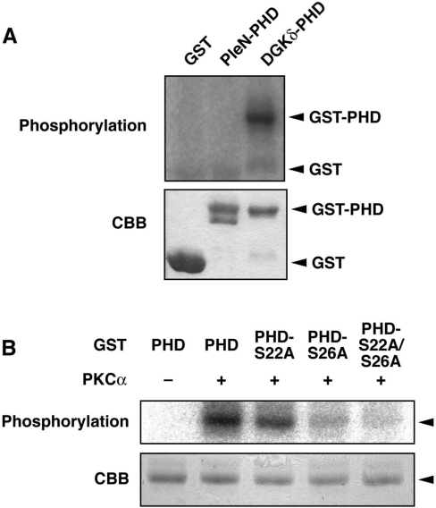
Figure 4. In vitro phosphorylation of the DGKδ1-PH domain by PKCα.
(A) GST, GST–pleckstrin N-terminal PH domain (PleN-PHD) and GST–DGKδ1-PH domain (DGKδ-PHD) proteins were incubated with (+) or without (−) purified rat PKCα (>90% purity). (B) GST–DGKδ1-PH domain (PHD), GST–DGKδ1-PH domain-S22A (PHD-S22A), GST–DGKδ1-PH domain-S26A (PHD-S26A) or GST–DGKδ1-PH domain-S22A/S26A (PHD-S22A/S26A) proteins were incubated with (+) or without (−) PKCα. Subsequently, the phosphorylated products were separated by SDS/PAGE followed by Coomassie Brilliant Blue staining (CBB, lower panels). The radioactive signal was visualized by a BAS1800 Bio–Image Analyzer (upper panels). The position of the GST–DGKδ1-PH domain and its mutants is indicated by an arrowhead.

