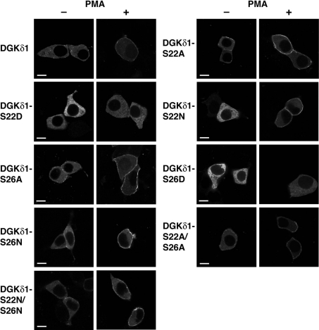
Figure 8. Effects of alanine/asparagine and aspartate mutations of Ser-22 and Ser-26 on the subcellular distribution of full-length DGKδ1.
HEK-293 cells were transfected with plasmids encoding YFP–DGKδ1 (DGKδ1), YFP–DGKδ1-S22A (DGKδ1-S22A), YFP–DGKδ1-S22D (DGKδ1-S22D), YFP–DGKδ1-S22N (DGKδ1-S22N), YFP–DGKδ1-S26A (DGKδ1-S26A), YFP–DGKδ1-S26D (DGKδ1-S26D), YFP–DGKδ1-S26N (DGKδ1-S26N), YFP–DGKδ1-S22A/S26A (DGKδ1-S22A/S26A) or YFP–DGKδ1-S22N/S26N (DGKδ1-S22N/S26N) as indicated. After starvation, cells were incubated for 30 min in the presence of 100 nM PMA (+) or 0.1% DMSO (−). After stimulation, HEK-293 cells were fixed with 3.7% (w/v) formaldehyde and then mounted on to glass slides. Cells were examined using an inverted confocal laser scanning microscopy (Zeiss LSM 510). Scale bar, 10 μm. Representatives of three repeated experiments are shown.

