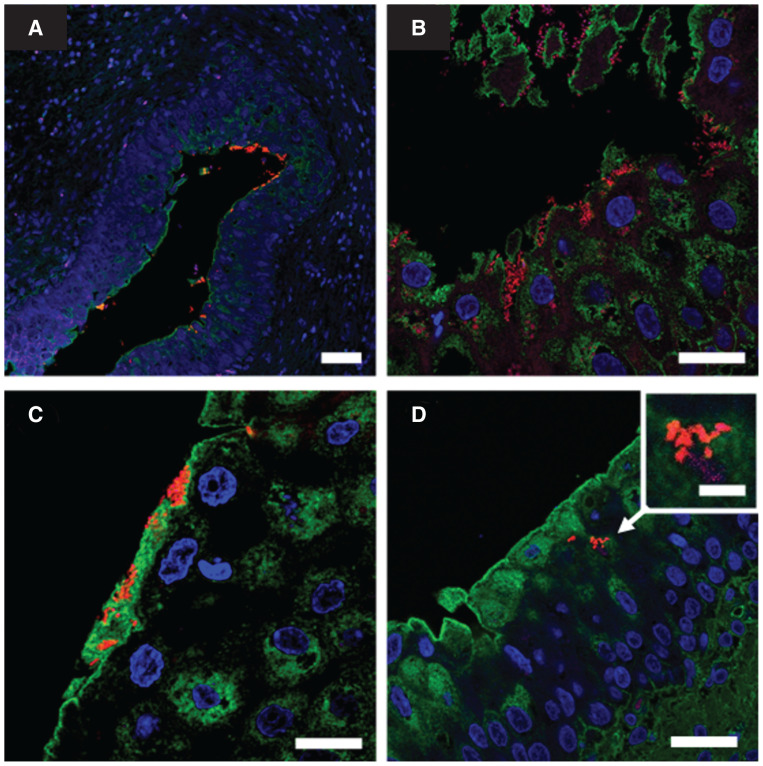
FIGURE 2.
Fluorescent in situ hybridization (FISH) analysis of bladder wall biopsies from a female dog. A — Patchy bacterial aggregates adhering to urothelium are visible at low magnification (scale bar = 50 μm). B — Invasive bacterial aggregates are observed in an area of urothelium erosion (scale bar = 25 μm). C — Bacterial aggregates embedded in a polysaccharide-rich matrix close to umbrella cells (scale bar = 25 μm). D — Image suggestive of bacterial aggregates within umbrella cells cytoplasm (scale bar = 25 μm); inset: intracellular bacterial community (scale bar = 5 μm). The FISH analysis stains are as follows: blue, 4′,6-diamidino-2-phenylindole (nuclear stain); green, wheat germ agglutinin (polysaccharide-rich material stain); red, Eub338 universal probe (bacterial stain).

