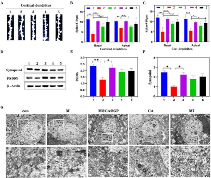
Figure 4.
Reprinted with permission from ref (136). Copyright 2022 Elsevier. (A) The study includes representative images of dendritic segments from cortical layer II/III pyramidal neurons, visualized using Golgi staining on brain sections. (B, C) Quantitative analysis was conducted to determine the density of apical and basal dendritic segments in cortical layer II/III pyramidal neurons, as well as spine density in hippocampus CA1 pyramidal neurons. (D) Western blotting analysis was performed to detect specific protein expression levels. (E, F) The study involved quantifying the levels of Synapsin1 and PSD95 proteins in the cortex, providing insights into synaptic protein dynamics. (G) Representative images obtained through Transmission Electron Microscopy (TEM) illustrate the ultrastructure of dorsal hippocampus neurons. The experimental conditions or treatments mentioned (M, RBC-MIC hybrid membrane; CA, curcumin-aspirin esterl; MI, minocycline; MCAGP, CA loading the graphene oxide quantum dots nanosize carrier with the modification of a hybrid cell membrane) were applied to investigate their effects on the observed parameters, such as dendritic morphology and protein levels.

