Figure 1.
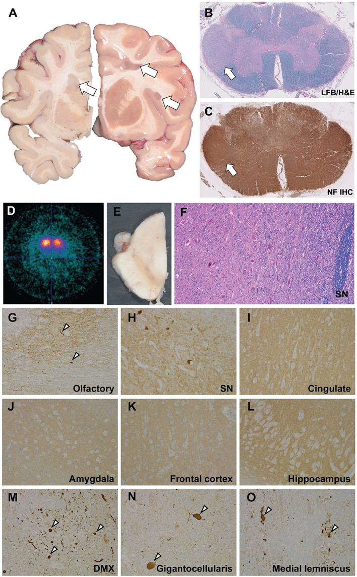
Neuropathological and neuroimaging evaluation demonstrating multiple sclerosis and brainstem degeneration in the absence of nigral Lewy bodies. (A) Gross coronal sections showing scattered white matter plaques (white arrows). (B) Luxol fast blue counterstained hematoxyoin & eosin stained (LH&E) section from the cervical spinal cord showing myelin pallor in the lateral funiculus. (C) Immunohistochemistry (IHC) using antisera targeting neurofilament shows intact axons consistent with a demyelinating process. (D) b-CIT SPECT revealed marked, symmetrically reduced uptake indicating significant loss of striatal dopamine neuronal innervation. (E) Gross hemisection of the midbrain demonstrating marked pallor in the substantia nigra. (F) High power (20x) photomicrograph of an LH&E-stained section through the midbrain showing marked loss of pigmented dopaminergic neurons and reactive gliosis. Immunohistochemistry to α-synuclein shows mild Lewy pathology (white arrowheads) in the olfactory bulb (G), negative staining in the substantia nigra (H), cingulate gyrus (I), amygdala (J), frontal cortex (K), hippocampus (L), and positive staining in the medulla, including the dorsal motor nucleus of the vagus (DMX; M), nucleus gigantocellularis (N), and medial lemniscus (O).

