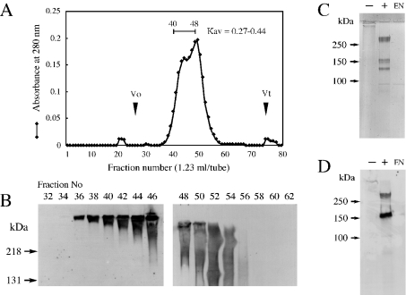
Figure 1. Purification and identification of syndecan-3-binding protein.
(A) Syndecan-3-binding protein was fractionated by chromatography on Sepharose CL-4B and the elution profile and elution volume Ve were analysed. The void volume Vo and the maximum elution volume Vt were measured with respect to the elution positions of Blue Dextran and p-nitrophenol respectively. (B) Aliquots of each fraction were subjected to ligand overlay assay using biotinylated soluble syndecan-3. (C, D) Positive fractions [Kav=(Ve−Vo)/(Vt−Vo)=0.27–0.44] were collected, treated with (+) or without (–) chondroitinase ABC and then separated by SDS/PAGE (6% gel), followed by either visualization by CBB staining (C) or immunochemical detection using anti-neurocan monoclonal antibody 3A11 (D). EN, the same amount of chondroitinase ABC used in (C, D) was subjected to SDS/PAGE followed by the same procedure as described above.

