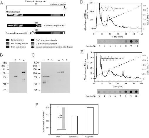
Figure 6. Binding of recombinant N- and C-terminal neurocan fragments to heparin or HSPGs.
(A) FLAG-tagged recombinant neurocan fragments, as represented schematically, were prepared as described in the Materials and methods section. EGF, epidermal growth factor; GAG, glycosaminoglycan; HA, hyaluronic acid; Ig, immunoglobulin. (B) BL21 competent cells were transformed using plasmids constructed with cDNA encoding the N-terminal (lanes 1 and 2) or C-terminal fragment (lanes 3 and 4) of mouse neurocan. Before (lanes 1 and 3) and after (lanes 2 and 4) induction with isopropyl β-D-thiogalactoside, lysates of each type of cell were subjected to SDS/PAGE (7% gel) followed by immunoblot analysis. FLAG-tagged recombinant neurocan fragments were detected with HRP-conjugated anti-FLAG monoclonal antibodies. (C) Cell lysates containing either the FLAG-tagged recombinant N-terminal (lanes 1–3) or C-terminal (lanes 4–6) recombinant neurocan fragment were mixed with anti-FLAG monoclonal antibody-conjugated agarose (lanes 1 and 4), heparin–Sepharose (lanes 2 and 5) or Sepharose CL-4B (lanes 3 and 6). The bound proteins were subjected to SDS/PAGE and immunoblot analysis as described above. Additional bands corresponding to lower molecular masses, which were detected in (B) (lane 4) and (C) (lanes 1 and 2), were probably produced by proteolysis. (D, E) Cell lysates containing the FLAG-tagged recombinant N- (D) or C-terminal (E) neurocan fragment were applied to a column of TSKgel Heparin-5PW. The bound proteins were eluted with a linear gradient of NaCl from 0.1 to 1.0 M. FLAG-tagged recombinant neurocan fragments were detected by dot-blot analysis with HRP-conjugated anti-FLAG monoclonal antibodies. (F) Purified N-terminal neurocan fragment was subjected to SDS/PAGE and stained with Coomassie Brilliant Blue (inset). This fragment was added to the wells coated with syndecan-3 or glypican-1. Bound fragment was detected as described in the Materials and methods section.

