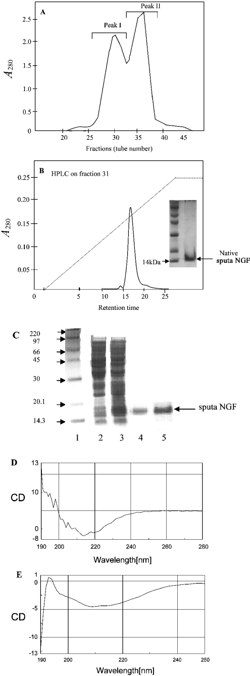
Figure 1. Purification of NGF.
(A) Gel-filtration chromatography of crude N. sputatrix venom. (B) Reversed-phase chromatography of the active fraction obtained from gel filtration. Inset shows the protein (native sputa NGF fraction) analysed by SDS/PAGE. (C) Purification and refolding of recombinant NGF (sputa NGF). Lane 1, protein molecular mass standards (sizes indicated in kDa); lane 2, E. coli cell lysate before induction; lane 3, E. coli cell lysate (inclusion bodies) after 3 h induction; lane 4, purified sputa NGF; lane 5, refolded sputa NGF. (D) CD spectrum obtained from refolded recombinant sputa NGF and (E) CD spectrum of native NGF protein.

