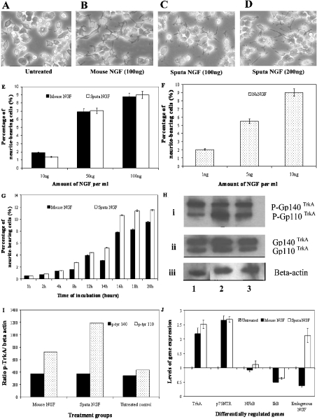
Figure 3. Neurite extension and receptor protein expression after treatment with mouse and sputa NGF.
PC12 cells at 1×10⁶ were plated and grown overnight. On the following day, cells were treated with either mouse or sputa NGF, at 200 ng for 16–18 h unless otherwise stated. (A, B) Neurite extensions observed after treatment with mouse NGF. (C, D) Neurites induced by sputa NGF. (E) Neurite extensions quantified for mouse (black bars) and sputa (white bars) NGF (10, 50 and 100 ng). (F) Neurite outgrowth observed for native nsNGF (stippled bars) at 1, 5 and 10 ng. (G) Time-course induction of neurite outgrowth by mouse (black bars) and sputa (white bars) NGF. (H) Activation of TrkA receptor determined by Western-blot analysis. Cells were treated for 15 min and total protein was extracted. Equal amounts of protein were separated on a SDS/7.5% polyacrylamide gel and subsequently blotted on to nitrocellulose membrane. (i) Phosphorylated TrkA receptors (gp110 and gp140) were detected using monoclonal anti-phosphotyrosine antibody. The same membrane was stripped and probed with (ii) anti-TrkA and (iii) β-actin antibodies. Lane 1, mouse NGF-treated cells; lane 2, sputa NGF-treated cells and lane 3, untreated control cells. (I) Ratios of the phosphorylated TrkA and β-actin (internal control) levels measured by densitometry. (J) Quantitative gene analysis via SYBR Green assay. The RNAs from treated and untreated samples were used to study the expression of five genes: TrkA, p75NTR, NF-κB and IκB.

