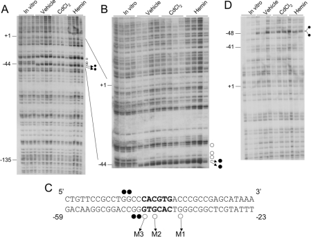
Figure 1. In vivo DMS footprinting of the human HO-1 proximal promoter in HK-2 cells.
(A) The transcribed strand of the human HO-1 promoter. Each lane is derived from an individual plate of HK-2 cells treated with vehicle (DMSO), CdCl2 (cadmium chloride, 10 μM) or haemin (5 μM) for 2 h. In vitro samples are derived from DNA purified from uninduced cells and then subsequently treated as described in the Experimental section. ○, Protected guanine residues; •, enhanced guanine residues. −135, −44 and +1 represent nucleotide positions relative to the transcription start site of the human HO-1 gene. (B) The same samples shown in (A) were electrophoresed for a longer run to visualize the E-box and distal sequences more closely. (C) The sequence of the human HO-1 proximal E-box (in bold) and flanking nucleotides. The guanine residues protected and enhanced in (A, B) are shown. The three protected guanine residues were assigned as M1, M2 and M3 and represent the sites for mutagenesis. (D) In vivo DMS footprinting of the non-transcribed strand of HO-1. •, Enhanced guanine residues at positions −47 and −48 bp as depicted in (C).

