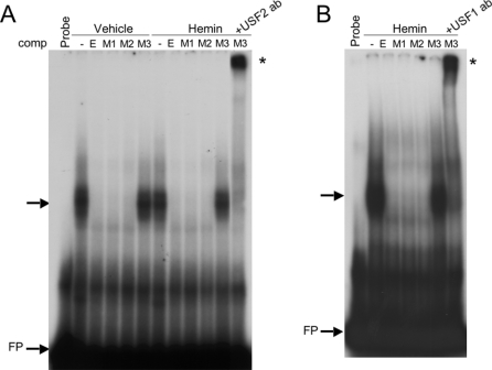
Figure 5. Importance of the guanine at −44 bp for DNA–protein binding.
(A) EMSA using the HO-1 proximal E-box with either vehicle- or haemin (5 μM)-treated nuclear extracts. Shifted DNA–protein complexes are indicated by an arrow, supershifted complexes are indicated with *. Unlabelled competitors are: E, E-box; M1, M2 and M3 represent G→A mutations of the three DMS-protected G bases shown in Figures 1(A) and 1(B). USF2 antibody was added after the addition of the unlabelled M3 competitor. (B) EMSA as seen in (A) using anti-USF1 antibody for supershift analysis. FP, free probe.

