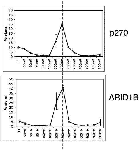
Figure 8. DNA-binding affinity of p270 and ARID1B.
In vitro translated [35S]methionine-labelled peptides were applied to native DNA–cellulose columns as described in the Materials and methods section. Bound protein was eluted stepwise with loading buffer adjusted to contain increasing concentrations of NaCl from 100 to 800 mM, as indicated in the Figure. Fractions were separated by SDS/PAGE and the p270 signal in each fraction was quantified by phosphoimaging. The results are plotted as the percentage of signal in each fraction relative to the entire signal recovered. Error bars represent S.D. Graphs are aligned for ease of comparison.

