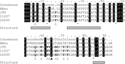
Figure 1. Amino acid sequence alignment of HU from H. pylori J99, 11637 and 26695.
The consensus sequence, derived from alignment of 60 HU homologues (with positions of >80% homology identified; [27]), is shown at the top, followed by the sequence of HBsu. Residues are numbered based on the H. pylori HU sequences. Residues that vary between HpyHU J99, 11637 and 26695 sequences are identified with an asterisk. Residues that correspond to the overall consensus sequence are highlighted in black, and HpyHU-specific divergent residues are identified with an open triangle. The DNA-intercalating Pro64 is identified by the closed triangle. Helical segments, based on the structure of B. stearothermophilus HU, are indicated by hatched boxes below the alignment.

